
Imaging Fibrous Structure Abnormalities of the White of the Eye in Myopathic Patients
Researchers provide an innovative approach to understanding ocular pathologies by visualizing the fiber structure of the sclera, the outermost eye layer
Tokyo, Japan – Eye diseases are extremely prevalent worldwide, with recent estimates suggesting that one-third of the global population suffers from some type of vision impairment. Given the high complexity of the human eye, the precise origin and nature of many eye diseases remain unclear, leaving affected people with limited diagnostic and treatment options.
Now, in a study made available online on March 7, 2024 and published in Volume 142, Number 4 of JAMA Ophthalmology on April 1, 2024, a team of researchers from Tokyo Medical and Dental University (TMDU) in Japan have pioneered the use of a novel kind of optical coherence tomography (OCT), a technique otherwise widely used in clinical ophthalmology, to investigate the detailed structure of the sclera—the white outer layer of the eyeball.
The motivation underlying this work stems from the limited options currently available to ophthalmologists for investigating the finer details of the sclera in living patients and specimens. “The sclera, composed of collagen fibers, plays an important role in protecting the retina, optic nerve, and other nerve tissues in the eye. Therefore, abnormalities in the shape of the sclera can cause various complications leading to blindness,” explains lead author Dr. Kyoko Ohno-Matsui. “Until now, however, the sclera of live subjects has only been measured in terms of its thickness, with no way to obtain details such as the orientation of the collagen fibers over a wide area of the eye.”
To overcome this limitation, the researchers developed a setup to conduct polarization-sensitive OCT (PS-OCT), a technique where the polarization of light acts as a contrast mechanism. “The sclera has a property called birefringence, which is an optical property of materials in which the refractive index depends on polarization. Birefringence is typically observed in fibrous tissues that have periodically organized nanostructures, such as the sclera,” comments senior author Dr. Tae Igarashi-Yokoi. “Thus, in addition to the magnitude of birefringence, which gives us information about the density of fibers, PS-OCT can also show the axis of orientation of the birefringence, which is related to the orientation of the fiber bundles themselves.”
Using this technique, the team investigated the properties of the collagen fibers in the sclera of patients with highly myopic eyes. They also focused on the link between myopathy and a sometimes-pathological condition known as dome-shaped macula (DSM), in which a specialized area in the retina bulges outwards. Their analysis included 89 highly myopic eyes from a total of 72 patients, mostly adults over 50 years old.
After careful observation of the acquired PS-OCT images, the researchers found that the sclera is divided into inner and outer layers with different structural arrangement for each. In the inner layer, the fibers extend radially from the periphery of the optic nerve. In contrast, fibers in the outer layer run perpendicularly to those of the inner layer. Interestingly, in patients with DSM, the fibers of the inner layer were aggregated and thickened, whereas those of the outer layer were compressed and thinned.
The successful use of PS-OCT to visualize the organization of fibrous tissue in eye structures could have huge implications for clinical research, diagnostics, and therapeutics. “Given the common occurrence of scleral pathologies, such as DSM and staphylomas in eyes with myopia, recognizing fiber patterns could provide important insights that may be relevant to developing targeted therapies to address scleral abnormalities early and mitigate potential damage to the overlying neural tissue,” remarks senior author Dr. Masahiro Yamanari.
We certainly hope PS-OCT leads to impactful medical discoveries that will enable more people to protect their treasured sense of vision.
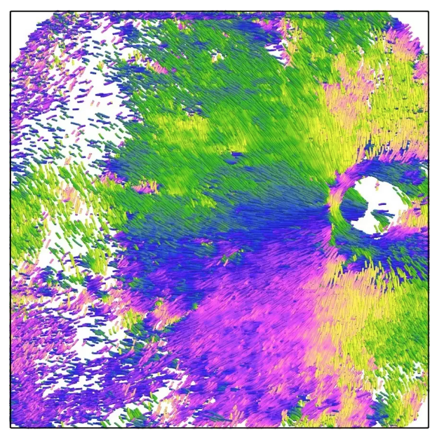
Visualization of the orientation and density of structural fibers in the sclera, the outermost eye layer
Researchers from Japan successfully applied polarization-sensitive optical coherence tomography (PS-OCT) to assess various properties of the collagen fibers that make up the sclera, the outer white of the eye. PS-OCT could determine the direction of these fibers at different layers of the sclera, enabling researchers to compare these features between eyes with different ocular pathologies.
###
The article, “Polarization-Sensitive OCT Imaging of Scleral Abnormalities in Eyes with High Myopia and Dome-Shaped Macula,” was published in JAMA Ophthalmology at DOI: 10.1001/jamaophthalmol.2024.0002
Summary
Journal Article
TITLE:Polarization-Sensitive OCT Imaging of Scleral Abnormalities in Eyes With High Myopia and Dome-Shaped Macula
DOI:https://doi.org/10.1001/jamaophthalmol.2024.0002
Correspondence to
Kyoko Ohno, Professor
Department of Ophthalmology and Visual Science,
Graduate School of Medical and Dental Sciences,
Tokyo Medical and Dental University(TMDU)
E-mail:k.ohno.oph[@]tmd.ac.jp
*Please change (at) in e-mail addresses to @ on sending your e-mail to contact personnels.