“Discovery of a novel function of dendritic cells to fine-tune excessive immune responses”

“Discovery of a novel function of dendritic cells to fine-tune excessive immune responses”

Toshiaki OHTEKI
Professor, Department of Biodefense Research,
Medical Research Institute

– A new avenue of treatment for infectious and autoimmune disease –

Highlights

1.An immune response is a double-edged sword that simultaneously defends and injures the host; the more severe the infection, the greater the regulatory control must be.
2.Our research group discovered that hemophagocytosis by dendritic cells moderates immune responses to ensure the host’s survival.
3.Our findings may lead to hemophagocytosis-based diagnostic and therapeutic applications for viral infections and autoimmune diseases.
Our research group, led by Prof. Toshiaki Ohteki at the Department of Biodefense Research, Medical Research Institute, Tokyo Medical and Dental University, in collaboration with Prof. Kenichi Sawada at the Department of Hematology, Nephrology, and Rheumatology, Akita University Graduate School of Medicine, has discovered a novel function of dendritic cells (DCs) for fine-tuning excessive immune responses in vivo. Under steady-state conditions, DCs consist of conventional DCs (cDCs) and plasmacytoid DCs (pDCs). DCs may also be derived from inflammatory monocytes (monocyte-derived DCs, Mo-DCs), especially under inflammatory conditions. The present study demonstrated that, during severe viral infections, Mo-DCs engulfed apoptotic erythroid cells in a process called hemophagocytosis, which is a characteristic of hemophagocytic syndrome (HPS). Importantly, hemophagocytosis was required for Mo-DCs to produce interleukin-10 (IL-10), an important immunoregulatory cytokine, thereby fine-tuning the immune responses to limit self-damage and ensure the host’s survival. These findings shed light on the physiological relevance of hemophagocytosis in severe inflammatory and infectious diseases, and may assist in identifying valuable therapeutic targets for severe inflammatory diseases. The study was supported by a research grant from JST, CREST, and was published online in Immunity on September 12, 2013 (EST).

Main findings and significance of this study

Hemophagocytic syndrome (HPS), which is characterized by fever, hepatosplenomegaly, cytopenia, hypertriglyceridemia and/or hypofibrinogenemia, and hemophagocytosis, is a severe, often fatal inflammatory disease. HPS is generally divided into primary and acquired forms. Our research group initially found that high doses of Toll-like receptor (TLR) ligands, such as CpG and poly I:C, when injected into wild-type (WT) mice, produced symptoms of HPS and induced hemophagocytosis, which is typically defined as the engulfment of erythroid cells by Mo-DCs in the blood, spleen, and bone marrow (Fig.1, left panel). Most viruses that induce HPS in humans establish a chronic infection. Thus, to examine the cellular and molecular events underlying hemophagocytosis, we used the lymphocytic choriomeningitis virus (LCMV) variant clone 13 (C13), which also elicits a chronic infection in mice. In addition, LCMV C13 infects DCs with high affinity and proliferates vigorously, possibly mimicking the effect of high-dose CpG and poly I:C injection. As expected, LCMV C13 infection effectively induced hemophagocytosis in WT mice (Fig. 1, right panel). In this context, LCMV infection-induced type I interferons (IFNs) were necessary for both the erythroid cell expression of the apoptosis indicator phosphatidylserine (PS), and the Mo-DC expression of the PS receptors. Importantly, injecting antibodies (Abs) against the Mo-DC PS receptor, which blocks hemophagocytosis, effectively reduced production of the regulatory cytokines, IL-10 and TGF-β1 in LCMV C13-infected WT mice (Fig. 2). This finding suggested that the Mo-DC production of IL-10 in response to C13 infection is hemophagocytosis-dependent. To examine the physiological relevance of the hemophagocyte-derived IL-10 in viral infection, Cd11c-cre Il10fl/fl (CKO) mice, in which the hemophagocytes cannot produce IL-10, were infected with LCMV C13. Importantly, the CKO mice showed excessive cytotoxic T lymphocyte (CTL) activity, tissue damage, and mortality (Fig. 3). Taken together, these results point to hemophagocytosis as a mechamism that ensures the host’s survival by preventing excessive immune response-mediated damage, instead allowing the virus to persist in the host under conditions of severe viral infection.

In summary, our findings indicate that hemophagocytosis is induced by TLR ligands or viruses in sequential steps (Fig. 4) to suppress potentially damaging immune responses.

Figure

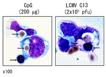
Figure 1. Hemophagocytosis by Mo-DCs.
Sorted hemophagocytes from the bone marrow of CpG-injected or LCMV C13-infected mice were stained with Diff-Quick. Black arrows indicate hemophagocytosed and attached erythroid cells. Original magnification, x100.

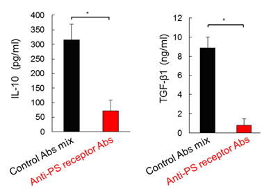
Figure 2. Hemophagocytosis-dependent IL-10 and TGF-β1 production.
Amount of serum IL-10 and TGF-β1 24 h in WT mice after LCMV C13 infection, which was injected with an isotype-control Ab cocktails or Abs that block PS receptors (500 μg each).

Figure 3. Hemophagocytosis fine-tunes immune responses in viral infection.
Amount of serum aspartate aminotransferase (AST) and alanine aminotransferase (ALT), indicators of liver damage, in WT and CKO mice 9 days after infection with LCMV C13, and the survival (%) of WT or CKO mice after infection.

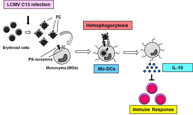
Figure 4. The development and role of hemophagocytosis in immune responses.
Type I IFNs produced in response to LCMV C13 infection induce phosphatidylserine (PS) on erythroid cells and PS receptors on Mo-DCs. Mo-DCs perform hemophagocytosis and release the immunoregulatory cytokine IL-10 to fine-tune immune responses, including CTL activity.

Correspondence to:

Toshiaki OHTEKI, Ph.D.
Department of Biodefense Research
Medical Research Institute, Tokyo Medical and Dental University
1-5-45 Yushima, Bunkyo-ku, Tokyo 113-8510, Japan
E-mail: ohteki.bre(at)mri.tmd.ac.jp

*Please change (at) in the e-mail addresses to @ when sending correspondence.