
Breaking through barriers
Tokyo, Japan – During pregnancy, the human placenta plays multiple essential roles, including hormone production and nutrient/waste processing. It also serves as a barrier to protect the developing fetus from external toxic substances. However, the placental barrier can still be breached by certain drugs. In a recent article published in Nature Communications, a team led by researchers at Tokyo Medical and Dental University (TMDU) developed a trophoblast stem (TS) cell-based organoid model of the placental barrier to support further biological research.
Villi in the human placenta help form the barrier and are surrounded by a layer of cells called trophoblasts. Because the structural nature of villi is critical for its function, cell lines and other methods used to replicate placental physiology in laboratory experiments have proven inadequate. Primary placental cells are also difficult to maintain in culture. Therefore, the TMDU group aimed to develop an effective in vitro model of placental villi using TS cells.
“TS cells have the capacity to differentiate into all kinds of placental cells consisting of the human placenta.,” says Dr. Takeshi Hori, lead author of the study. “However, it has been challenging to make the barrier model using TS cells.”
First of all, the team then generated trophoblast organoids, a type of three-dimensional cell model that can more effectively mimic the structural and biological details of an organ. After testing three types of culture medium, they determined the optimal conditions to support the formation of spherical organoids.
“The outer layer of the organoid contained a single layer of cells called syncytiotrophoblasts,” explains Dr. Hirokazu Kaji, senior author. “This layer effectively displayed the barrier function that we were aiming to mimic with this model.”
Based on the culture conditions of the spherical organoids, the researchers established flatter organoids with a column-type container to easily asses the translocation of compounds through the barrier layer. The researchers used various methods to confirm the barrier integrity and maturation levels of the plane organoids and to ensure the robustness of the system. Their analysis also showed that the model could be used to assess how well different compounds could cross the barrier, specifically by examining the permeability coefficients.
“Using the organoids as a model of the placental barrier will help scientists better understand general placental biology and potential drug toxicity,” says Dr. Hori. “We also designed our model in a manner such that the cells could be easily cultured and it could be evaluated using microscopic observation.”
The TS cell-based organoid model generated in this study effectively addresses many of the difficulties that have previously hampered laboratory-based assessments of placental physiology. It will be a useful tool for not only elucidating details of the development of this organ, but also for evaluating the transfer rates and toxicity levels of various compounds. This will be critical in the drug development process to avoid damaging the placenta or harming the fetus.
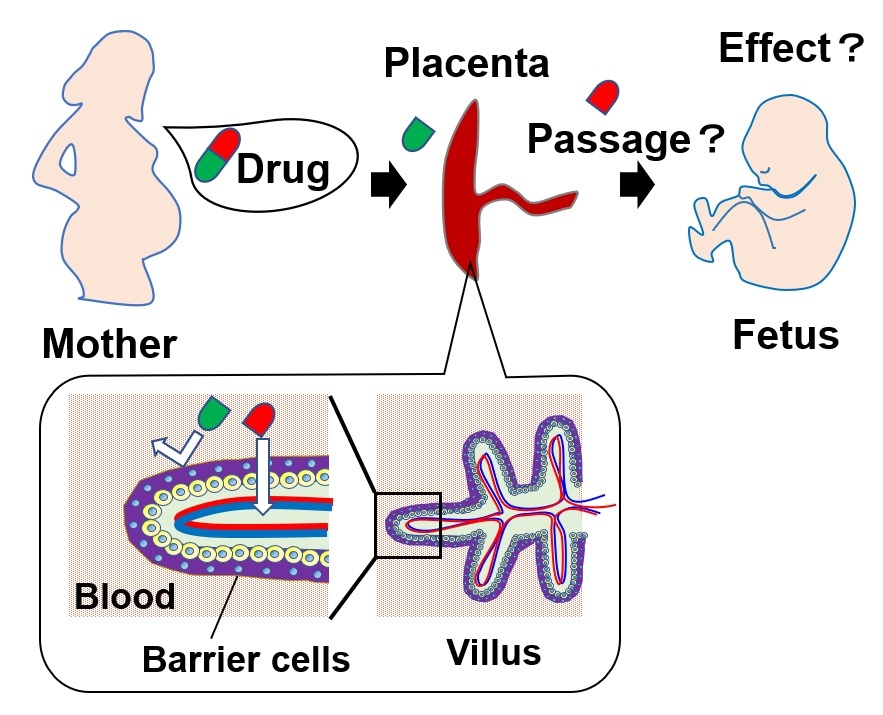
Figure 1. Some drugs can cross the placental barrier and reach the fetus
In the human placenta, there are placental villi, and the surface of the villi consists of syncytiotrophoblast, also called barrier cells, that serve as the main barrier against foreign substances. However, some medicines taken by pregnant women can penetrate this placental barrier and have undesirable effects on the fetus.
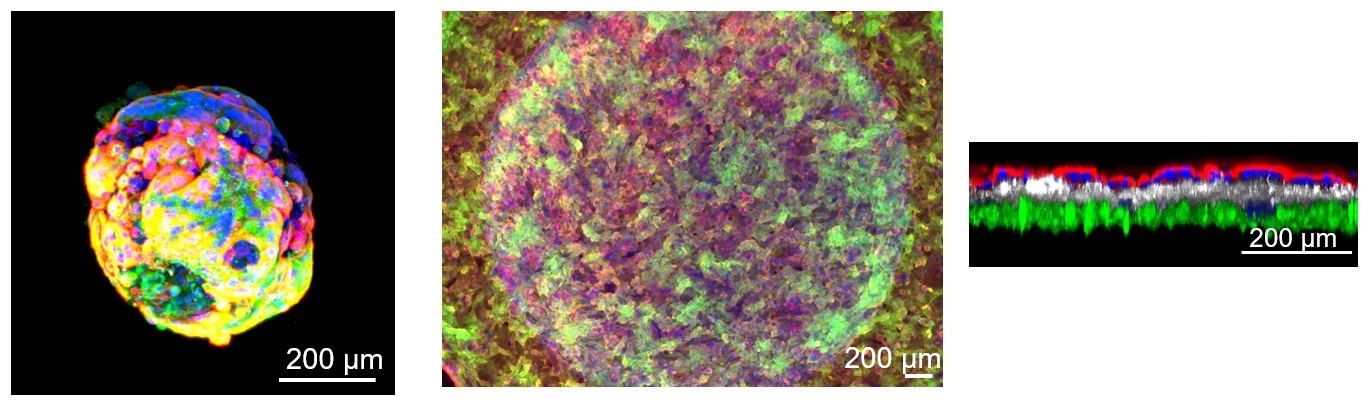
Figure 2. Placental organoids and barrier models
Placental organoids generated from human placental stem cells (left), a front view of the placental barrier model (center), and a side view of the barrier model (right image, red-colored cells at the top of the layers indicate barrier cells).
The article, “Trophoblast stem cell-based organoid models of the human placental barrier,” was published in Nature Communications at DOI: 10.1038/s41467-024-45279-y
Summary
Journal Article
TITLE:Trophoblast stem cell-based organoid models of the human placental barrier
DOI:https://doi.org/10.1038/s41467-024-45279-y
Correspondence to
Hirokazu Kaji, Professor
Department of Diagnostic and Therapeutic Systems Engineering,
Institute of Biomaterials and Bioengineering,
Tokyo Medical and Dental University(TMDU)
E-mail: kaji.bmc(at)tmd.ac.jp
*Please change (at) in e-mail addresses to @ on sending your e-mail to contact personnels.