
“Injected Mix of Bone-augmenting Agents Causes New Bone Growth in Mouse Jaws”
Researchers centered at Tokyo Medical and Dental University(TMDU) deliver a protein/peptide combination using an injectable gel carrier to promote bone formation in mouse jawbones
Tokyo – The part of the jawbone containing tooth sockets is known as alveolar bone, and its loss over time or following dental disease may ultimately result in tooth loss. While dentures can be used as a tooth replacement, the mechanical stimuli under the dentures causes further bone loss. An alternative and more permanent solution is strongly hoped for. Recombinant human bone morphogenetic protein 2 (BMP-2) has been used to stimulate osteogenesis (bone formation) in humans, but high levels can cause inflammation and tumor development. Therefore, agents such as peptide drugs for accelerating bone augmentation need to be developed, even in the presence of lower levels of BMP-2. Additionally, there are no known means of stimulating local bone augmentation without performing surgery.
The peptide OP3-4 has been shown to inhibit bone decay and stimulate the differentiation of cells (osteoblasts) that form bone. Now, an international team centered at Tokyo Medical and Dental University has injected a gelatin-based gel carrying OP3-4 and BMP-2 into mice jawbones to trigger local augmentation of bone around the injection site. The study was recently reported in the Journal of Dental Research.
Use of this injectable gelatin-based gel to carry the agents avoids the need for surgical implantation and resulted in no swelling or other such complications in the experimental mice. The researchers observed a region of increased bone mass around the BMP-2 + OP3-4 injection site that was larger than that seen in mice injected with BMP-2 alone, or with other controls. This mass also had a significantly higher bone mineral content and density (Image1).
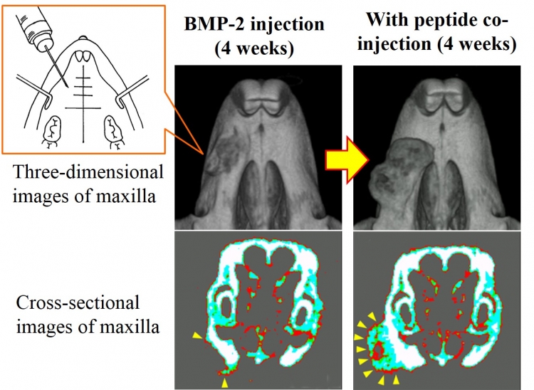
Image 1: Peptide co-injection thickens mouse bone
Yellow arrowheads indicate newly formed bone
Microscopic examination revealed the deposition of calcified tissue (mineralization) throughout the newly formed bone of BMP-2 + OP3-4-treated mice.
“Mineralization of the outer region evidently took place before that of the inner region,” lead author Tomoki Uehara says (Image2). “We speculate that the size of the new bone is determined before calcification starts, and that OP3-4 plays an important role in making a regeneration site at the early stage of bone formation.”
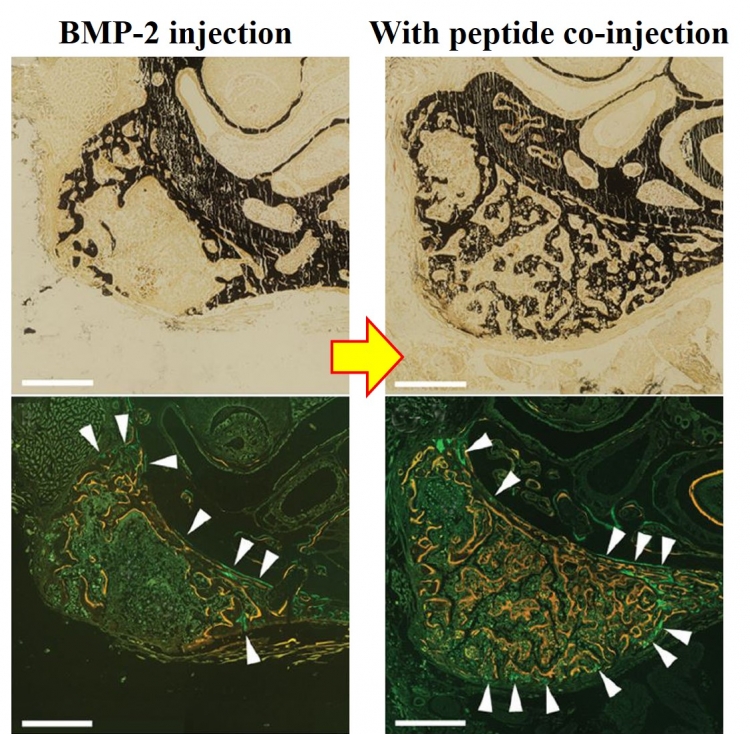
Image 2: Microscopic views of newly formed bone
Black area mainly shows newly formed bone (upper panel). Green lines (white arrowheads) and yellow lines indicate bone formation activity on day 12 and 26 after injections, respectively. Bar represent 0.5 mm.
Corresponding author Kazuhiro Aoki adds: “OP3-4 further enhanced the number of bone-forming cells induced by BMP-2 treatment, and greatly increased the expression of genetic markers of bone formation.”
The article “Delivery of RANKL-Binding Peptide OP3-4 Promotes BMP-2-Induced Maxillary Bone Regeneration” was published in the Journal of Dental Research at DOI: 10.1177/0022034516633170.
Summary
Correspondence to:
Department of Bio-Matrix (Pharmacology),
Graduate School of Medical and Dental Sciences,
Tokyo Medical and Dental University (TMDU)
Phone:+81-3-5803-5461
E-mail: kazu.hpha(at)tmd.ac.jp