
Transistors help identify cancer cell markers
Researchers from Tokyo Medical and Dental University (TMDU) find that ion-sensitive field effect transistors can be combined with enzymatic chemical signal amplification to detect a cancer-related marker in breast cancer cells
Tokyo, Japan - Having biopsies taken and endless tests run is nobody’s idea of a good time, even if it’s necessary for monitoring your health. Now, researchers from Japan report the development of a new technique that could make testing for cancer a lot less invasive.
In a study published in September in the Journal of the American Chemical Society, researchers from Tokyo Medical and Dental University (TMDU) have revealed a new technique for assessing the cancer-related marker using breast cancer cell lines.
Detecting cancer-related markers is a powerful way to determine diagnosis, prognosis, and treatment success. Modern techniques are capable of detecting these markers in patient samples such as blood and urine, which provides a noninvasive way to monitor and evaluate patients.
“Circulating tumor cells (CTCs), which are cancer cells found in the blood, are one of the main targets used to evaluate cancer patients’ blood samples,” states Miyuki Tabata, first author of the study. “However, it can be challenging to isolate these cells from the blood, and current approaches do not adequately detect both epithelial cell and mesenchymal cell markers, which are important for determining the stage of the cancer.”
To create a system that can quickly and easily detect cancer-related markers on CTCs (and potentially other factors in the blood), the researchers used an apparatus called an ion-sensitive field effect transistor (ISFET), which is a tiny electrical circuit that is activated by a change in pH. They coated these transistors with breast cancer cells and then added an antibody linked to a chemical reporter that causes a change in pH if the antibody recognizes the cells.
“We found that the chemical reporter glucose oxidase successfully detected the expression of epidermal growth factor receptor (EGFR), a marker of poor cancer prognosis, on CTC membranes,” says Yuji Miyahara, senior author of the study. “Furthermore, the strength of the chemical signal correlated with the amount of EGFR expressed by the cells.”
Importantly, these results corresponded to the levels of EGFR detected using other techniques, indicating that the ISFET approach accurately identifies cancer-related marker expression on cells.
“These results provide proof of concept that an ISFET-based system can be used to efficiently assess patient cancer status based on liquid biopsy samples,” states Tabata.
Given that ISFETs can be manufactured to be the size of a single cell and assembled into vast arrays, this technique has the potential to enable high-throughput analysis of cancer cells at single-cell resolution. In addition, the use of multiple antibodies for the chemical enzyme detection step could enable the simultaneous analysis of multiple cancer-related markers.
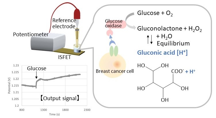
Detection principle of membrane proteins using the cell based-transistor
Ion-sensitive field effect transistors detect breast cancer cells expressing EGFR when bound by an antibody linked to a chemical reporter
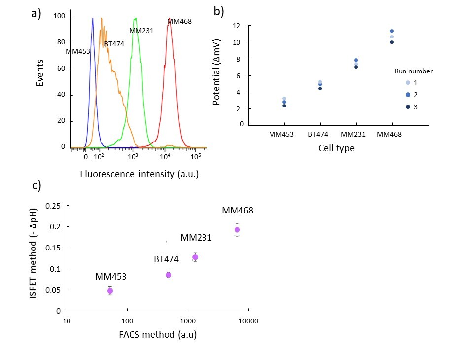
Comparison of membrane protein expression measurement between the conventional method and the developed cell based-transistor
a) Flow cytometry results in four types of breast cancer cell lines
b) Cell-transistor results in four types of breast cancer cell lines
c) Correlation between flow cytometry results and cell-transistor results
The results of membrane protein measurement using the cell-transistor correlate with the results of conventional fluorescence-activated cell sorting (FACS). Realization of an ultra-sensitive liquid biopsy platform based on semiconductor technology is expected.
The article, “Detection of epidermal growth factor receptor expression in breast cancer cell lines using an ion-sensitive field effect transistor in combination with enzymatic chemical signal amplification,” was published in the Journal of the American Chemical Society at DOI: 10.1021/jacs.2c06122
Summary
Journal Article
TITLE:Detection of epidermal growth factor receptor expression in breast cancer cell lines using an ion-sensitive field effect transistor in combination with enzymatic chemical signal amplification
DOI:https://doi.org/10.1021/jacs.2c06122
Correspondence to
Miyuki Tabata, Specially Appointed Associate Professor
Yuji Miyahara,Specially Appointed Professor
Department of Bioelectronics,
Institute of Biomaterials and Bioengineering,
Tokyo Medical and Dental University(TMDU)
E-mail:miyahara.bsr(at)tmd.ac.jp
*Please change (at) in e-mail addresses to @ on sending your e-mail to contact personnels.