
A new polarized fluorescent probe for revealing architectural dynamics of living cells

Researchers from Tokyo Medical and Dental University (TMDU), collaborating with scientists from the Marine Biological Laboratory (MBL) and RIKEN, develop a novel technique for live-cell fluorescent imaging which leads them to discover a new actin structure in starfish early embryos.
Tokyo, Japan – Monitoring alignments of the building blocks of cells is important to understand how the cells are built. By collaborating with imaging scientists at the MBL, researchers from Japan have developed a new probe which they call POLArIS, allowing real-time imaging of molecular orientations in live cells.
A fluorophore emits polarized light as it glows. The orientation of polarized fluorescence is closely related to the orientation of the fluorophore. If a molecule of interest is rigidly connected with a fluorescent tag such as Green Fluorescent Protein (GFP), the polarized fluorescence from the fluorophore reports the orientation of the molecule.
“In previous approaches for monitoring the orientation of the protein of interest, researchers needed to develop effective constrained GFP tagging methods which might be different for each protein of interest,” says one of the lead authors of the study, Ayana Sugizaki. “POLArIS uses an antibody-like binder that is rigidly connected with GFP, allowing both specific and versatile constrained labeling,” adds another lead author Keisuke Sato. The team used a commercially available Adhiron molecule (now rebranded as “Affimer”) as the binder molecule to link GFP to a target protein, and developed POLArIS by connecting Adhiron and GFP in a rotationally constrained manner (Figure 1). Because an Adhiron molecule that specifically binds to a molecule of interest can be easily selected from a library of molecules through phage display screening, POLArIS can be designed for any biological molecules of interest. POLArIS can be expressed in specific cell types and organelles, and will be useful for studying architectural dynamics of molecular assemblies in broad range of cell cultures, tissues and whole organisms. “From the point of view of fluorescence polarization imaging, POLArIS has significant advantages because of its genetically encoded nature,” says Tomomi Tani, a Senior Researcher at the National Institute of Advanced Industrial Science and Technology (AIST) in Japan, who has joined this project since he was an Associate Scientist at the MBL.
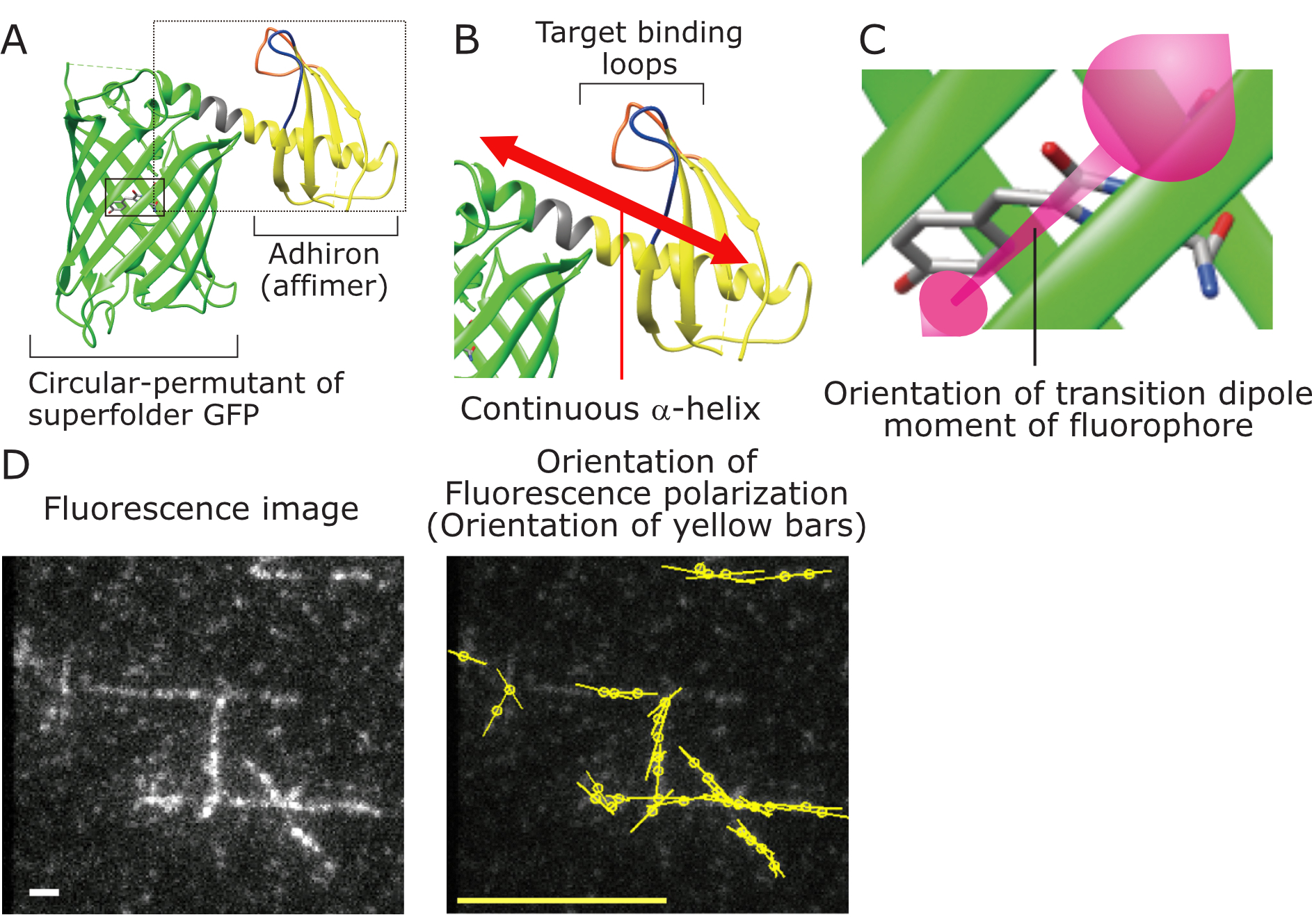
Figure 1
A. Overview of POLArIS structure. Green, Circular-permutant of superfolder GFP (cpGFP); Yellow, Adhiron (Affimer); Gray, α-helical linker connecting cpGFP and Adhiron; Blue and orange, Target binding loops.
B. Enlarged picture of the region of dashed-line box in A. cpGFP and Adhiron are rigidly connected by an α-helical linker.
C. Enlarged picture of the region of solid-line box in A. The orientation of fluorescence polarization of POLArIS reflects the orientation of transition dipole moment of fluorophore indicated by the magenta double-headed arrow.
D. Purified POLArIS protein that specifically binds to F-actin was added to actin filaments reconstituted in vitro, and was observed with fluorescence polarization microscopy. Left: Fluorescence image. White scale bar, 1 μm. Right: Polarization state of fluorescence spots in the left picture was analyzed. Orientation and length of yellow bars indicate the orientation and the strength of fluorescence polarization, respectively. Yellow scale bar, polarization factor=0.5.
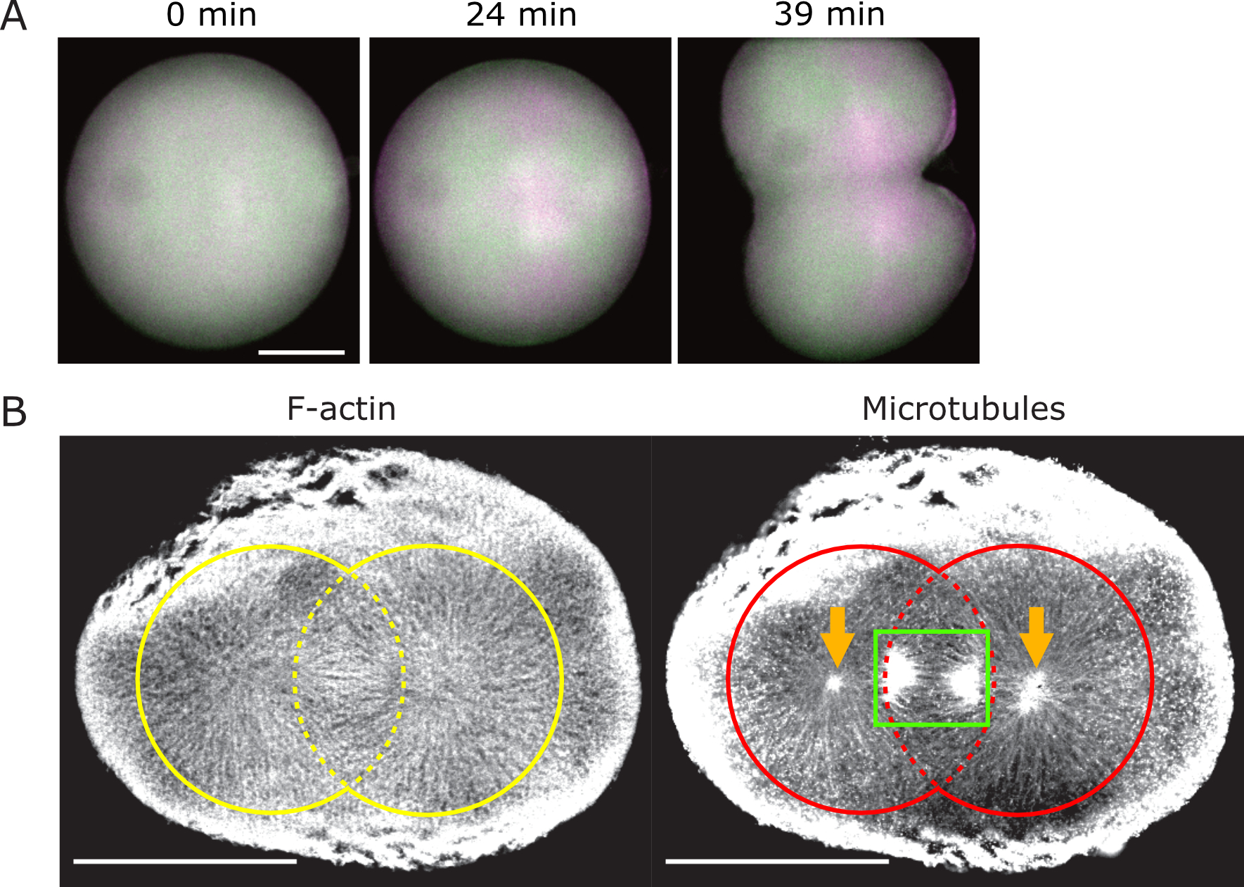
Figure 2
A. Live-cell fluorescence polarization imaging of the first cleavage of a starfish embryo expressing POLArIS that specifically binds to F-actin. Vertical and horizontal actin filaments are shown in magenta and green, respectively. FLARE structures made of radially extending actin filaments were observed as cross-patterns of magenta and green. Scale bar, 50μm.
B. A starfish embryo in the first cleavage was fixed, stained with fluorescent-dye-conjugated phalloidin (stains F-actin) and anti-tubulin antibody (stains microtubules), and observed with confocal laser scanning microscope. Yellow circles, regions of FLARE structure; Red circles, regions of astral microtubules; green box, mitotic spindle; orange arrows, centrosomes. Scale bar, 100 μm.
Time-lapse movies of the polarized fluorescence imaging during the first cleavage of a starfish embryo expressing POLArIS that specifically binds to F-actin in a rotationally constrained manner (the same egg as in Fig. 2A). Magenta parts in the left movie indicate horizontally aligned actin filaments and the green parts for those of actin filaments that are vertically aligned. Right, the same movie with enhanced contrast.
Summary
Journal Article
Proceedings of the National Academy of Sciences of the United States of America
TITLE:
POLArIS, a versatile probe for molecular orientation, revealed actin filaments associated with microtubule asters in early embryos
DOI:
https://doi.org/10.1073/pnas.2019071118
Correspondence to
Department of Neuroanatomy and Cellular Neurobiology
Graduate School of Medical and Dental Sciences,
Tokyo Medical and Dental University(TMDU)
1-5-45 Yushima, Bunkyo-ku, Tokyo 113-8510, Japan
E-mail:terada.nana(at)tmd.ac.jp
*Please change (at) in e-mail addresses to @ on sending your e-mail to contact personnels.