“Identification of a novel mechanism by which allergic inflammation is dampened”

“Identification of a novel mechanism by which allergic inflammation is dampened”

Hajime KARASUYAMA
Professor
Department of Immune Regulation,
Graduate School of Medical and Dental Sciences (right)

Mayumi EGAWA
Graduate Student
Department of Immune Regulation,
Graduate School of Medical and Dental Sciences (left)

- Converting immune cells from being proinflammatory to anti-inflammatory -
A group led by Prof. Hajime Karasuyama at Department of Immune Regulation, Tokyo Medical and Dental University, Graduate School of Medical and Dental Sciences has discovered a novel mechanism by which proinflammatory cells are converted to anti-inflammatory cells, leading to the suppression and termination of allergic skin inflammation in a mouse model of atopic dermatitis. The failure of this conversion resulted in exacerbation and prolongation of allergic inflammation in the skin. This discovery would prompt the identification of novel therapeutic targets for the treatment of allergic disorders. The study was carried out in collaboration with Prof. Naofumi Mukaida at Cancer Research Institute, Kanazawa University, and supported by a research grant from JST, CREST. The research results were published online in Immunity on February 22nd 2013.

Highlights

1.As many as ~30% of people in Japan suffer from allergic disorders including allergic rhinitis, atopic dermatitis, and asthma.
2.The mechanism for the development of allergy has been extensively studied while that for the suppression and termination of allergy has been poorly understood.
3.The present study identified a novel mechanism by which allergic inflammation is dampened, via conversion of inflammation-inducing (proinflammatory) cells to inflammation-suppressing (anti-inflammatory) cells.
4.This finding would promote the development of a novel strategy for the treatment of allergic disorders.

Main findings and significance of this study

Karasuyamas’ group has previously developed the mouse model of IgE-mediated, chronic skin inflammation that mimics human atopic dermatitis. Many white blood cells (leukocytes) are recruited to and accumulated in the allergen-exposed skin, leading to the swelling and redness of the skin. The present study demonstrated that inflammatory monocytes represented the major population among leukocytes recruited to the skin lesions (Figure 1). Inflammatory monocytes express a chemokine receptor CCR2 that is essential for their migration into peripheral tissues such as the skin. Therefore, it was expected that mice lacking CCR2 might show reduced allergic inflammation in the skin, due to the impaired recruitment of inflammatory monocytes to the skin lesions. In contrast to this expectation, the skin allergy was severer and prolonged in these mice in spite of the absence of inflammatory monocytes in the skin lesions. Injection of CCR2-expressing inflammatory monocytes isolated from normal mice into CCR2-deficient mice dampened the exacerbated allergic inflammation in the skin of CCR2-deficient mice (Figure 2), indicating that inflammatory monocytes exerted an anti-inflammatory function in this allergic reaction. Previous studies showed that inflammatory monocytes differentiate into type 1 macrophages after their migration into peripheral tissues, and induce inflammation (Figure 3). The present study illustrated that inflammatory monocytes recruited to the allergic skin differentiated into type 2 macrophages, under the influence of a cytokine (interleukin-4) produced by basophils, and dampened allergic inflammation (Figure 1). Two modes of type 2 macrophage generation have been reported: one is the differentiation from blood-circulating, resident monocytes, and the other is the differentiation from tissue-resident macrophages (Figure 3). The present study identified the third mode of type 2 macrophage generation, namely, the differentiation from blood-circulating, inflammatory monocytes. Further elucidation of molecular mechanisms underlying this differentiation and the anti-inflammatory action of type 2 macrophages would promote the development of a novel strategy for the treatment of allergic disorders.

Figures

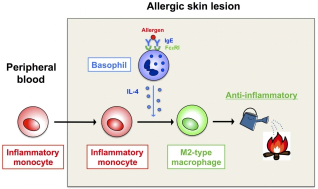
Figure 1. Conversion of immune cells from being proinflammatory (inflammatory monocytes) to anti-inflammatory (type 2 macrophages)
Inflammatory monocytes recruited to the allergen-exposed skin differentiate into type 2 macrophages, under the influence of a cytokine (interleukin-4) produced by basophils, and dampen allergic inflammation.

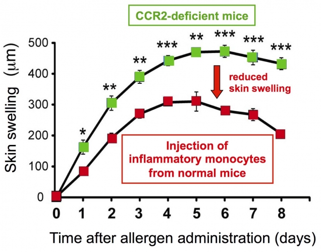
Figure 2. Inflammatory monocytes show anti-inflammatory property after their migration to allergic skin
Mice lacking a chemokine receptor CCR2 show severer and prolonged skin swelling due to impaired migration of inflammatory monocytes to the skin lesions. Injection of CCR2-expressing inflammatory monocytes isolated from normal mice into CCR2-deficient mice dampens the exacerbated allergic inflammation in the skin of CCR2-deficient mice.

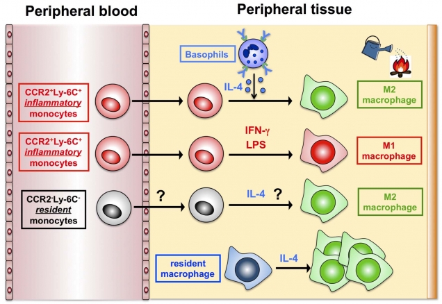
Figure 3. A novel mode of type 2 macrophage generation
It is well known that blood-circulating, inflammatory monocytes differentiate into type 1 macrophages that show an inflammatory property, after their recruitment to peripheral tissues. In contrast, the mode of type 2 macrophage generation has been poorly understood. The present study identified a novel mode of type 2 macrophage generation, that is, the differentiation from blood-circulating, inflammatory monocytes, besides previously-reported modes: the generation from blood-circulating, resident monocytes and that from tissue-resident macrophages.

Correspondence to:

Hajime KARASUYAMA, MD, PhD.
Department of Immune Regulation
Tokyo Medical and Dental University, Graduate School of Medical and Dental Sciences
1-5-45 Yushima, Bunkyo-ku, Tokyo, Japan 113-8519

Phone: +81-3-5803-5162
E-mail: karasuyama.mbch(at)tmd.ac.jp


URL: http://immune-regulation.org/index.php?id=27

*Please change (at) in e-mail addresses to @ upon sending your e-mail to the contact personnel.