“Development of non-human primate model of amyotrophic lateral sclerosis (ALS)”

“Development of non-human primate model of amyotrophic lateral sclerosis (ALS)”

Takanori Yokota
Professor,
Department of Neurology and Neurological Science,
Graduate School of Medical and Dental Sciences (right)

Hidehiro Mizusawa
Chairman,
Department of Neurology and Neurological Science,
Graduate School of Medical and Dental Sciences (left)

A research group led by Professor Takanori Yokota and Chairman Hidehiro Mizusawa at Department of Neurology and Neurological Science, Graduate School of Medical and Dental Sciences, Tokyo Medical and Dental University developed a non-human primate model of amyotrophic lateral sclerosis (ALS). This was achieved in cooperation with the research groups of Tsukuba Primate Research Center, National Institute of Biomedical Innovation, Tokyo Metropolitan Institute of Medical Science, and Chiba University. This work was supported by Comprehensive Research on Disability Health and Welfare; Research on Neurodegenerative Diseases/ALS from Ministry of Health, Labor and Welfare, Japan; Grant-in-Aid for Scientific Research (A) and Grant-in-Aid for Research Activity Start-up and Strategic Research Program for Brain Science, Field E from Ministry of Education, Culture, Sports and Technology, Japan. The research results were published online on 18, Jan 2012 in Brain.

Point

We first developed a non-human primate model of ALS with cytoplasmic mislocalization of TDP-43.
There is a species difference in TDP-43 pathology, and our monkey model recapitulates ALS pathology to a much greater extent than rodent models, providing a valuable tool for studying the pathogenesis and developing therapy of sporadic ALS.

Content of the research

Amyotrophic lateral sclerosis (ALS) is a fatal neurodegenerative disease characterized by progressive motoneuron loss. Redistribution of TDP-43 from the nucleus to the cytoplasm is considered pathological hallmarks of amyotrophic lateral sclerosis, but their significance has not been fully elucidated. Since all reported rodent transgenic models using wild-type TDP-43 failed to recapitulate these features, we expected a species difference and aimed to make a non-human primate model of ALS. We overexpressed wild-type human TDP-43 in spinal cords of cynomolgus monkeys and rats by injecting adeno-associated virus vector into the cervical cord, and examined the phenotype using behavioural, electrophysiological, neuropathological and biochemical analyses. These monkeys developed progressive motor weakness and muscle atrophy with fasciculation in distal hand muscles first (Fig. A). They also showed regional cytoplasmic TDP-43 mislocalization with loss of nuclear TDP-43 staining in the lateral nuclear group of spinal cord innervating distal hand muscles (Fig. B and C) and the Betz cell in the primary motor cortex (Fig. D), reminiscent of the spinal cord pathology of ALS patients. TDP-43 mislocalization was an early or presymptomatic event and was later associated with neuron loss. These findings suggest that the TDP-43 mislocalization may lead to motoneuron degeneration. Furthermore, truncation of TDP-43 was not a prerequisite for motoneuronal degeneration, and phosphorylation of TDP-43 occurred after degeneration had begun. In contrast, similarly prepared rat models expressed TDP-43 only in the nucleus of motoneurons. There is thus a species difference in TDP-43 pathology, and our monkey model recapitulates ALS pathology to a greater extent than rodent models, providing a valuable tool for studying the pathogenesis and developing therapy of sporadic ALS.

Contact

Takanori Yokota, MD, PhD
Department of Neurology and Neurological Science,
Tokyo Medical and Dental University
1-5-45 Yushima Bunkyo-ku, Toko 113-8519
Phone: +81-3-5803-5234
FAX: +81-3-5803-0169
email: tak-yokota.nuro(at)tmd.ac.jp
*Please change (at) in e-mail addresses to @ on sending your e-mail to contact personnels.

Figure

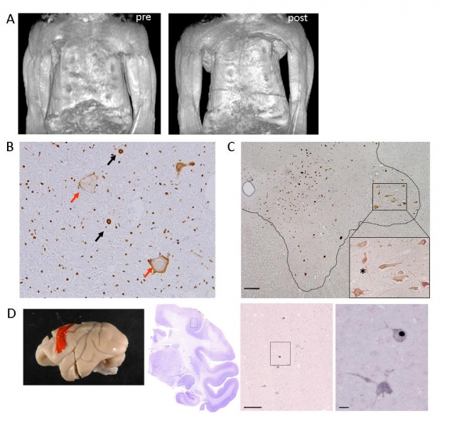
Overexpressing wild-type TDP-43 in the monkey cervical cord leads to progressive motor weakness and muscle atrophy. Motor paresis of the forelimb on the injected side (encircled) in TDP-43 AAV-injected monkeys. (A) Constructed MRI images of upper arm muscles before (pre) and 4 weeks after (post) injection, indicating a marked muscle atrophy of upper and forearms as well as hand muscles, predominantly on the injected side (left side). (B) Immunostaining of the cervical cord sections of TDP-43–injected monkeys at the late stage using antibodies to pan-TDP-43 demonstrated normal localization in nuclei (black arrows), mislocalization in cytoplasm (red arrows). Regional mislocalization of TDP-43 and cell death in monkey spinal cords. (C) Sections from the injected side of the 8th cervical segment of the cord taken at the early stage and immunostained with an anti-Flag antibody. Most neurons in the lateral nuclear group (area encircled by broken line) showed cytoplasmic mislocalization of TDP-43 (enlarged inset), but almost all neurons in other areas expressed exogenous TDP-43 in the nucleus.(D) Immunostaining with anti-Flag antibody indicated that exogenous flag TDP-43 is expressed in nucleus or cytoplasm of the Betz cells in the primary motor cortex. Scale bars: 200 μm.