
New imaging technique for faster and more accurate detection of cavities
PDF Download
- Accurate detection of cavities

Junji Tagami
Executive Director at TMDU
Executive Vice President of Education and
International Student Exchange
Professor of Cariology and Operative Dentistry
Profile
Dr. Tagami completed his dental and graduate school at TMDU, where he received his DDS and PhD. He became Adjunct Assistant Professor at the Medical College of Georgia at Augusta University, and in 1994, assumed the post of Professor and Chairman of the Department of Operative Dentistry at Ohu University. He returned to TMDU in 1995 as Professor and Chairman of Cariology and Operative Dentistry. Dr. Tagami became Executive Director and Executive Vice President in 2014.
Please tell us about this new technology.
A: The method we employ is called sweptsource optical coherence tomography (SS-OCT), which uses high-speed frequency swept-laser light from a near-infrared laser. The light is projected at the occlusal surface of the tooth and scanned across its proximal surface. Twodimensional images from below the tooth surface are generated by detecting the backscattered laser beam signal, which is digitized over a time scale.
A: These days, oral health professionals typically use radiographs to detect cavities that are not visible to the naked eye. However, cavitated enamel lesions and dentin caries that comprise cavities can be difficult to detect on radiographic images, especially at the early stages. Our study has shown that images generated using SS-OCT are better than radiographic images for detecting cavities in several ways. First, cavities were detected at an earlier stage with SS-OCT than with radiographs. Also, SS-OCT detected cavities in areas that may be difficult to reach using radiographs. Finally, SS-OCT appears to provide more reliable and accurate images than bitewing radiographs. In addition to these differences, SS-OCT images can be collected in ealtime, enabling on-site diagnosis in a dental clinic.
A: Earlier detection of cavities using SS-OCT would allow the cavity to be treated when it is smaller, rather than detecting it later when the cavity could be larger and require more extensive and invasive treatment. This could reduce the pain experienced by the patient, which is associated with more complex dental procedures. Moreover, SS-OCT imaging does not require any radiation, so it can be used safely for dental diagnosis on patients such as pregnant woman and young children.
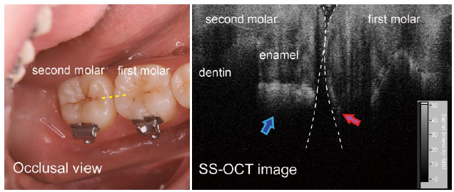
Occlusal view (left) and SS-OCT image (right) of lower first and second molars
The SS-OCT image was obtained by scanning along the dotted yellow line shown in the lefthand image. SS-OCT reveals slight demineralization at the first molar (red arrow) and enamel caries at the second molar (blue arrow).
A: We already saw an improvement in cavity detection using two-dimensional imaging. Obtaining three-dimensional images in real time will further improve detection of earlystage cavities and cavities in areas that may be difficult to reach using X-rays. Our goal is to improve technology that can be used in dental offices, and this research is important in improving overall oral care.
Journal Information
J. Biophotonics, doi: 10.1002/jbio.201200210