Microendodontics
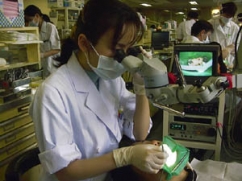
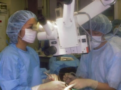
Stereomicroscope came to be used in the medical area for the first time in the 1950s, is now used in ophthalmology and neurosurgery in the 1960s. Use of stereomicroscope began to spread, particularly in Europe and the United States in the field of endodontics from 1990s. This device had been introduced to the surgical endodontic therapy initially, but it is also used in general root canal treatment.
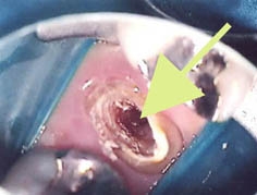
After access cavity prearation, when you remove the fracture device or post, and when you repair the perforated part, you should use the stereomicroscope. You can check the morphology of pulp chamber and root canal under a stereoscopic microscope, and it is easy to identify the remnants of the pulp chamber, and to discover the overlooked root canal, by the different color of dentin.
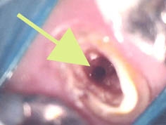
When treating the maxillary first molar, there are many overlooked mesial buccal second root canal (MB2), it causes the case such as "don't disappear the pain". Root canal, such as a mesial buccal second root canal does not always branche from the floor of pulp chamber, it sometimes branches from the middle of a root canal. After the root canal preparation, and observed under a stereoscopic microscope again a root canal, you can find a new root canal which branches in the middle root canal.
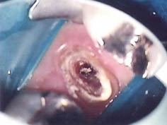
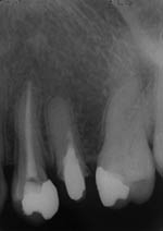
By using a stereo microscope, it is also possible to find out the calcified root canal . When you observe a stereoscopic microscope the root canal and pulp floor there, you can find easily root canal concealed. The above case would cut a direction different from the root canal with the naked eye, it is a case that you overlooked calcified root canal. Patience is need to find that fumbling root canal in the conventional method. However, We were able to look out the root canal of the original under a stereoscopic microscope, to perform the root canal filling in this case. It is able to confirm the broken line fracture of root canal using the stereomicroscope. After root canal preparation, If you look well under a stereoscopic microscope the root canal wall, you may want to find a micro-crack. It is important to care inside of canal walls in prosthesis to prevent spread the fracture.
The cases of retreatment, we sometimes found fractured equipment or post in the root canal. It is possible if fractured equipment and post exist in the near of orifice, it is neccessary to sacrifice the tooth more If you try to remove them. And it is hard to remove which had remained near the apex. It is possible to remove it by the combined use of ultrasonic device and stereoscopic microscope in such a case. Which might result in the root canal retreatment, perforation was observed in root canal orifice or near the bottom of pulp chamber. Under bright field at high magnification without causing overflow from the repairing material it is possible to verify the exact location of the device by using a stereomicroscope, and can be packed tightly to ensure the repairing materials.