A01-4
研究概要
光学顕微鏡・超解像光学顕微鏡を用いた細胞内小器官のイメージングを通じて、オルガネラゾーンの解明に貢献します。光学顕微鏡(マイクロスコープ)は、μmの現象の観察ツールです。近年、超解像光学顕微鏡が開発され、ナノメーターレベルの観察まで対象がひろがりました。超解像光学顕微鏡の応用の候補として、細胞内小器官の可視化解析が想定されています。一般の光学顕微鏡では観察が難しかったオルガネラも超解像顕微鏡を上手く使えば、数十-100nmの高解像度で観察可能です。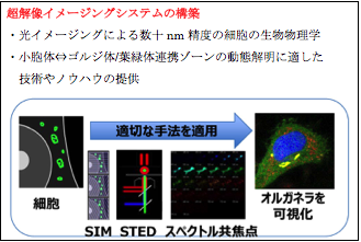
代表的な原著論文
-
Ohzono T.. Katoh K., Terentjev E.M. (2021)
Microscopy of diffuse nematic-isotropic transition in main-chain nematic liquid crystal elastomers. Macromolecules (in press) -
Ohzono T., Katoh K., Minamikawa H., Saed M. O., Terentjev E.M. (2021)
Internal constraints and arrested relaxation in main-chain nematic elastomers
Nat Commun 12, 787 (2021). https://doi.org/10.1038/s41467-021-21036-3 -
Higaki T., Akita K., Katoh K (2020)
Coefficient of variation as an image-intensity metric for cytoskeleton bundling
Sci. Rep. 10, 22187. https://doi.org/10.1038/s41598-020-79136-x -
Yamaguchi H., Honda S., Torii S., Shimizu K., Katoh K., Miyake K., Miyake N., Fujikake N.,
Sakurai H.T., Arakawa S., Shimizu S.(2020)
Wipi3 is essential for alternative autophagy and its loss causes neurodegeneration. Nat Commun 11, 5311. https://doi.org/10.1038/s41467-020-18892 -
Ueno Y., Matsuda K., Katoh K., Kuzuya A., Kakugo A., Konagaya A. (2020) Modeling a Microtubule Filaments Mesh Structure from Confocal Microscopy Imaging. Micromachines 11 (9), 844 https://doi.org/10.3390/mi11090844
-
Ishida K., Goto S., Ishimura M., Amanuma M., Hara Y., Suzuki R., Katoh K., Morita E. (2019)
Functional Correlation between Subcellular Localizations of Japanese Encephalitis Virus Capsid Protein and Virus Production.
J Virol 93:e00612-19. https://doi.org/10.1128/JVI.00612-19 -
Morita, M, Ota, Y, Katoh, K, Noda, N (2019) Bacterial Cell Culture at the Single-cell Level Inside Giant Vesicles.
JOVE-JOURNAL OF VISUALIZED EXPERIMENTS, (146):10.3791/59555 - Tanaka, M., Fujii, Y., Hirano, K., Higaki, T., Nagasaki, A., Ishikawa, R., Okajima, T., Katoh, K.(2019) Fascin in lamellipodia contributes to cell elasticity by controlling the orientation of filamentous actin. Genes to Cells. 24, p202-213, https://doi.org/10.1111/gtc.12671
- Takata, H., Madung, M., Katoh, K., Fukui, K.(2019) Cdk1-dependent phosphorylation of KIF4A at S1186 triggers lateral chromosome compaction during early mitosis. PLoS One 13(12): e0209614 doi.org/10.1371/journal.pone.0209614
- Morita, M., Katoh,K., Noda, N.(2018) Direct Observation of Bacterial Growth in Giant Unilamellar Vesicles: A Novel Tool for Bacterial Cultures. ChemistryOpen https://doi.org/10.1002/open.201800126
- Kijima, S. Staiger, C., Katoh, K., Nagasaki, A., Ito, K., and Uyeda, T. (2018) Arabidopsis vegetative actin isoforms, AtACT2 and AtACT7, generate distinct filament arrays in living plant cells. Sci Rep., 8:4381 | DOI:10.1038/s41598-018-22707-w.
- Ito, N., Katoh, K., Kushige, H., Saito, Y., Umemoto, T., Matsuzaki, Y., Kiyonari, H., Kobayashi, D., Soga, M., Era, T., Araki, N., Furuta, Y., Suda, T., Kida, Y., and Ohta K. (2018) Ribosome Incorporation into Somatic Cells Promotes Lineage Transdifferentiation towards Multipotency. Sci Rep. 8, 1634 | DOI:10.1038/s41598-018-20057-1
- Ohzono, T., Katoh, K., Wang, C., Fukazawa, A., Yamaguchi, S., and Fukuda, J. (2017) Uncovering different states of topological defects in schlieren textures of a nematic liquid crystal. Sci. Rep. 7, 16814 | DOI:10.1038/s41598-017-16967-1
- Nozumi, M., Nakatsu, F., Katoh, K., and Igarashi, M. (2017) The Coordinated Vesicle and Actin Bundle Movement in Nerve Growth Revealed by Superresolution Microscopy. Cell Rep. 18 2203–2216
- Ohzono, T., Katoh, K., and Fukuda, J. (2016) Fluorescence microscopy reveals molecular localisation at line defects in nematic liquid crystals. Sci. Rep. 6: 36477 | DOI: 10.1038/srep36477
- Shibata, T., Yamashita, S., Hirusaki, K., Katoh K., Ohta, Y. (2015) Isolation of mitochondria by gentle cell membrane disruption, and their subsequent characterization. Biochem. Biophys. Res. Commun. 463 563-568
- Sugiura, S., Nishimura, S., Yasuda, S., Hosoya, Y., and Katoh, K. (2006) Caron fiber technique for the investigation of single-cell mechanics in intact cardiac myocytes. Nat. Protoc. 1, 1453-1457
総説
- Pelc R., Hostounský Z., Otaki T., Katoh K. (2020) Conventional, Apodized, and Relief Phase-Contrast Microscopy. In: Pelc R., Walz W., Doucette J. (eds) Neurohistology and Imaging Techniques. Neuromethods, vol 153. Humana, New York, NY.
https://doi.org/10.1007/978-1-0716-0428-1_10 - 大瀧達朗、加藤薫(2018)位相差顕微鏡 実験医学 Vol36, No.20 (増刊) p24-25
- 加藤薫(2018)偏光観察 実験医学 Vol36, No.20 (増刊) p30-31
- 加藤薫(2018)複屈折顕微鏡(Polscope)実験医学 Vol36, No.20 (増刊) p73-75
- 大瀧達朗、加藤薫(2018)アポディぜーション位相差顕微鏡 実験医学 Vol36, No.20 (増刊) p80-81
- 加藤薫(2018) 超解像光学顕微鏡, 光技術動向調査報告書2017年度、p234-241
- 加藤薫、大瀧達朗 (2011) 新しいタイプの偏光顕微鏡とアポディゼーション位相差顕微鏡 O plus E 33, 274-278
- 金子浩子、杉浦清了、須田吉久、加藤薫 (2009) カーボン素材のバイオ分野への応用研究 化学工業 60, 66-73
- 加藤薫(2007) 光学顕微鏡を用いて微細な構造を見る −光による分子イメージング− 顕微鏡 42, 155-160
