Outline
Subjects of Interest
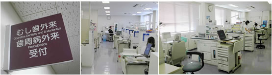
Laboratory Equipments
Recent Topics
Acid-base Resistant Zone
Fluorosed Tooth Bonding
Resin Coating Technique
Bonding to Root Dentin
Self Adhesive Resin Cements
Zirconia Bonding
Polymerization Shrinkage
Saliva Buffering
Biofilm Research
Micro-shear Bond Strength
Biocompatibility
Mechanical Behavior of Adhesives
Root Caries Prevention
Light Curing Unit
Optical Coherence Tomography
Our Research
Outline
Research at the Cariology and
Operative Dentistry involves a broad range of subjects ranging from the
basic science study on the caries process and to the development of
advanced clinical techniques in restorative dentistry. Main areas
include the evaluation of dental adhesives and other materials, studies
on oral biofilm and hard tissue remineralization, and development of aesthetic dental treatments
with regard to the "Minimal Intervention" concepts.
Subjects of Interest:
-Oral Biofilm
-Saliva Buffer Capacity
-Caries Mechanism
-Caries Inhibition
-Hard-tissue Remineralization
-Pulpal Healing
-Laser Treatment
-Ozone Treatment
-Effects of Fluoride
-Resin Coating Technique
-Interfacial Nanoleakage
-Acid-Base Resistant Zone
-Cavity Configuration Factor
-Durability of Adhesion
-Mechanical Characterization of Adhesives
-Micro-tensile Bond Strength
-Micro-shear Bond Strength
![]()
Laboratory Equipments:
-Scanning Electron Microscope (SEM)
-Optical and Stereo Microscopes
-Confocal Laser Scanning Microscope (CLSM)
-Argon Ion Etching
-Sputter Coater
-Thermo-cycling Device
-Low Speed Diamond Saws
-Microtome Saw
-Ion-selective Potentiometer
-Knoop Microhardness Tester
-Micro CT (Computed Tomography) System
-Nanoindentation System
-Universal Testing Machines (conventional and micro)
-Incubator
-pH meters (conventional and hand-held)
-Fluorescence Microscope
-Oral Biofilm Reactor (OBR)
-Lasers (CO2 and Er-YAG)
-Ozone Generator
![]()
Acid-base Resistant Zone
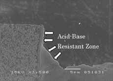 Good
adhesion is thought to enhance long-term sealing of the cavity margin,
resulting in protection of the restoration against secondary caries.
Artificial secondary caries around adhesive restorations placed in
bovine root dentin has been observed using a scanning electron
microscope (SEM). It was found that the change in ultrastructure of the
cavity margins after acid-base challenge was adhesive material
dependent. The hybrid layer was defined as a layer that the monomer
penetrated into the dentin and cured in situ. In order to distinguish
the hybrid layer dentin and resin, acid treatments, such as hydrochloric
acid and phosphoric acid, have been routinely used in previous studies.
Therefore, the hybrid layer was defined as an acid-base resistant zone
with respect to its characteristics against acid treatment. Using a
self-etching primer adhesive system, the so-called an “acid-base
resistant zone” was observed beneath the hybrid layer after acid-base
challenge. The adhesive resin had impregnated the exposed collagen
bundles and became entangled with them to create a hybrid layer, which
was distinguished by argon-ion etching and resistant to acid-base
challenge. Also, using different self-etching primer systems, an
acid-base resistant zone was detected after acid-base challenge, but
there was a difference morphologically and mechanically between the
systems.
Good
adhesion is thought to enhance long-term sealing of the cavity margin,
resulting in protection of the restoration against secondary caries.
Artificial secondary caries around adhesive restorations placed in
bovine root dentin has been observed using a scanning electron
microscope (SEM). It was found that the change in ultrastructure of the
cavity margins after acid-base challenge was adhesive material
dependent. The hybrid layer was defined as a layer that the monomer
penetrated into the dentin and cured in situ. In order to distinguish
the hybrid layer dentin and resin, acid treatments, such as hydrochloric
acid and phosphoric acid, have been routinely used in previous studies.
Therefore, the hybrid layer was defined as an acid-base resistant zone
with respect to its characteristics against acid treatment. Using a
self-etching primer adhesive system, the so-called an “acid-base
resistant zone” was observed beneath the hybrid layer after acid-base
challenge. The adhesive resin had impregnated the exposed collagen
bundles and became entangled with them to create a hybrid layer, which
was distinguished by argon-ion etching and resistant to acid-base
challenge. Also, using different self-etching primer systems, an
acid-base resistant zone was detected after acid-base challenge, but
there was a difference morphologically and mechanically between the
systems.
![]()
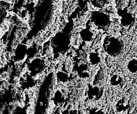
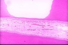
Dental fluorosis is a malformation of tooth enamel and dentin, which is
believed to be caused by chronic ingestion of fluoride during tooth
development. The structure of human fluorosed dentin shows
nanomorphological differences at the apatite crystallite and collagen
fibrillar level. These structural aberrations play a major role in
remarkable caries prone potential of the tissue, which is quite
obviously the opposite effect of fluorosed enamel. The currently
available adhesive systems do not offer the same bonding efficacy with
this pathological state of enamel and dentin tooth tissue.
Image: FE-SEM of fluorosed dentin treated with the enchant of an
adhesive system.
![]()
Resin Coating Technique
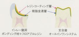 Tooth−colored
indirect restorations have become widely accepted. To overcome the
lackluster bonding performance to dentin, a resin coating technique was
developed. By means of a combined use of a dentin adhesive system and a
low-viscosity flowable resin composite or simply use of a dentin
adhesive system, the prepared dentin is immediately protected.
Therefore, this technique has the potential to minimize pulp irritation
and postoperative sensitivity and provide good bonding performance to
dentin. Further, resin coated dentin has resistance to caries because of
“Super Dentin” is formed on the dentin surface.
Tooth−colored
indirect restorations have become widely accepted. To overcome the
lackluster bonding performance to dentin, a resin coating technique was
developed. By means of a combined use of a dentin adhesive system and a
low-viscosity flowable resin composite or simply use of a dentin
adhesive system, the prepared dentin is immediately protected.
Therefore, this technique has the potential to minimize pulp irritation
and postoperative sensitivity and provide good bonding performance to
dentin. Further, resin coated dentin has resistance to caries because of
“Super Dentin” is formed on the dentin surface.
![]()
Bonding to Root Dentin
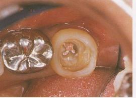 Composite
core systems can prevent root fracture of non-vital teeth; a good dentin
bonding system has been shown to reinforce the remaining tooth
structure. For successful adhesive restorations of non-vital teeth,
material selection is a key to obtaining good bonding to coronal, root
and pulpal floor dentin. In addition, it has been found that a
tight-sealing reduce the incidence of coronal leakage.
Composite
core systems can prevent root fracture of non-vital teeth; a good dentin
bonding system has been shown to reinforce the remaining tooth
structure. For successful adhesive restorations of non-vital teeth,
material selection is a key to obtaining good bonding to coronal, root
and pulpal floor dentin. In addition, it has been found that a
tight-sealing reduce the incidence of coronal leakage.
![]()
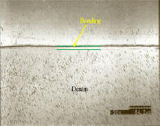
Self
Adhesive Resin Cements
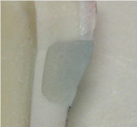 Self-adhesive
resin cements are the latest generation of materials in the adhesive
dentistry. These materials may simplify clinical procedures of
cementation and reduce the technique sensitivity of multi-step cement
systems. Those luting agents maybe applied in a single step and require
no pretreatment of the substrate dental tissue.
Self-adhesive
resin cements are the latest generation of materials in the adhesive
dentistry. These materials may simplify clinical procedures of
cementation and reduce the technique sensitivity of multi-step cement
systems. Those luting agents maybe applied in a single step and require
no pretreatment of the substrate dental tissue.
These dual-curing materials take advantage of the functional adhesive
monomer technology developed for self-etching primer adhesive systems.
Image: A self-adhesive resin cement has sealed the margins and the
dye penetration test showed no microleakage.
![]()
Zirconia Bonding
 Porcelain
fused to metal crowns and fixed partial dentures have been clinically
successful. Nevertheless, metal structures, even when covered with
ceramics, may represent an esthetic problem. On the other hand,
conventional ceramic materials used for all-ceramic restorations were
prone to failure due to their insufficient physical properties;
therefore, their use is limited for a single crown or short-span fixed
partial denture. Zirconia is used in biomedical applications and has
been recently introduced in restorative dentistry. The use of the
zirconium-oxide all ceramic material provides several advantages,
including biocompatibility, chemical stability, esthetic appearance and
high mechanical properties. Presently, zirconia is the only ceramic
material that has the potential to substitute the metal used in the
porcelain-fused-to-metal technology. Zirconia can be used for long-span
bridges. The only problem related to its performance is that adhesion of
resin cements to such ceramics is inferior. The long-term bonding
durability is inevitable for clinical success. Therefore, we work on the
bonding of resin cements to zirconia using veneering porcelain for
zirconia in order to obtain better bonding.
Porcelain
fused to metal crowns and fixed partial dentures have been clinically
successful. Nevertheless, metal structures, even when covered with
ceramics, may represent an esthetic problem. On the other hand,
conventional ceramic materials used for all-ceramic restorations were
prone to failure due to their insufficient physical properties;
therefore, their use is limited for a single crown or short-span fixed
partial denture. Zirconia is used in biomedical applications and has
been recently introduced in restorative dentistry. The use of the
zirconium-oxide all ceramic material provides several advantages,
including biocompatibility, chemical stability, esthetic appearance and
high mechanical properties. Presently, zirconia is the only ceramic
material that has the potential to substitute the metal used in the
porcelain-fused-to-metal technology. Zirconia can be used for long-span
bridges. The only problem related to its performance is that adhesion of
resin cements to such ceramics is inferior. The long-term bonding
durability is inevitable for clinical success. Therefore, we work on the
bonding of resin cements to zirconia using veneering porcelain for
zirconia in order to obtain better bonding.
![]()
Polymerization Shrinkage
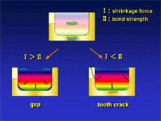 Light-cured
resin composites are now widely used in clinical practice on metal free
restorations, because of the tooth colored esthetic advantages. However,
the polymerization reactions of light-cured composites lead to
volumetric shrinkage. This contraction stress has been shown to lead to
marginal gaps, enamel and dentin cracks, postoperative sensitivity and
secondary caries. Recent research has shown it may be possible to reduce
the stresses within a bulk of resin composite using Slow-start curing
method, and flowable resin composite. Slow-start curing method can be
done, by prepolymerization at low light intensity followed by final cure
at high light intensity.
Light-cured
resin composites are now widely used in clinical practice on metal free
restorations, because of the tooth colored esthetic advantages. However,
the polymerization reactions of light-cured composites lead to
volumetric shrinkage. This contraction stress has been shown to lead to
marginal gaps, enamel and dentin cracks, postoperative sensitivity and
secondary caries. Recent research has shown it may be possible to reduce
the stresses within a bulk of resin composite using Slow-start curing
method, and flowable resin composite. Slow-start curing method can be
done, by prepolymerization at low light intensity followed by final cure
at high light intensity.
![]()
Saliva Buffering
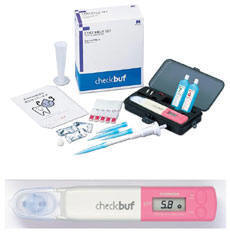 The
micro pH sensor was developed for the clinical usage with support by
HORIBA, Ltd. (Kyoto, Japan), and it is used for quantitative assessments
for caries activity and reminelarization process. We also developed the
quantitative saliva buffer capacity test using a hand-held pH meter (checkbuf®,
J Morita, Tokyo, Japan), and use it at the chair-side for caries risk
assessments.
The
micro pH sensor was developed for the clinical usage with support by
HORIBA, Ltd. (Kyoto, Japan), and it is used for quantitative assessments
for caries activity and reminelarization process. We also developed the
quantitative saliva buffer capacity test using a hand-held pH meter (checkbuf®,
J Morita, Tokyo, Japan), and use it at the chair-side for caries risk
assessments.
![]()
Biofilm Research
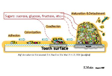 Oral
biofilm formation is a key factor in the process of caries. For
prevention of caries, in particular, new and effective prevention
methods were proposed including biofilm removal by alkaline water and
ozone disinfection of water.
Oral
biofilm formation is a key factor in the process of caries. For
prevention of caries, in particular, new and effective prevention
methods were proposed including biofilm removal by alkaline water and
ozone disinfection of water.
![]()
Micro-shear
Bond Strength
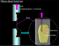 We
evaluate bonding performance and durability of adhesive materials using
newly developed bond test method. The new method enables to detect the
difference among the variation of tooth structure, and is widely
recognized as a precise bond test in dentistry. We also evaluate the
leakage of adhesive restorations under electron-microscopic level.
We
evaluate bonding performance and durability of adhesive materials using
newly developed bond test method. The new method enables to detect the
difference among the variation of tooth structure, and is widely
recognized as a precise bond test in dentistry. We also evaluate the
leakage of adhesive restorations under electron-microscopic level.
![]()
Biocompatibility
 We
evaluate biocompatibility and bioadaptability of restorative materials
using in vivo pulpal response test and so on. Especially, pulpal
response tests of composite resin restorations are continued and renewed
since the development of the material, of which results over many years
are recognized as valuable information in dental clinic.
We
evaluate biocompatibility and bioadaptability of restorative materials
using in vivo pulpal response test and so on. Especially, pulpal
response tests of composite resin restorations are continued and renewed
since the development of the material, of which results over many years
are recognized as valuable information in dental clinic.
![]()
Mechanical Behavior of Adhesives
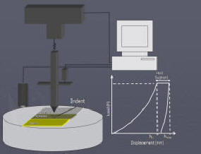 Dental
adhesive layer is a thin polymer film. Understanding and exploring the
mechanical properties of adhesive materials is necessary for advancement
in the adhesive dentistry. Besides the traditional mechanical testing
and informative bond strength tests, we have an interest in exploring
the specific mechanical behavior and relevant contributing factors of
dental adhesives, through alternative test techniques such as
nanoindentation.
Dental
adhesive layer is a thin polymer film. Understanding and exploring the
mechanical properties of adhesive materials is necessary for advancement
in the adhesive dentistry. Besides the traditional mechanical testing
and informative bond strength tests, we have an interest in exploring
the specific mechanical behavior and relevant contributing factors of
dental adhesives, through alternative test techniques such as
nanoindentation.
![]()
 The
prevention of root caries is the most important theme in aging society.
The root dentin is low resistant to caries. However, resistance to the
acid is given from a past research by spreading and using the bonding
material, and the prevention of the root caries. Resin coating technique
is available for inhibition of root dentin demineralization, however
prevention of root dentin demineralization varies in coating materials.
The
prevention of root caries is the most important theme in aging society.
The root dentin is low resistant to caries. However, resistance to the
acid is given from a past research by spreading and using the bonding
material, and the prevention of the root caries. Resin coating technique
is available for inhibition of root dentin demineralization, however
prevention of root dentin demineralization varies in coating materials.
![]()
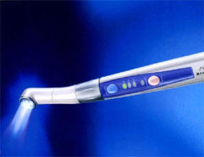
In the recent years, there has been an increasing
demand for aesthetic restorations. With the development of dental
bonding materials, cavities with various sizes and locations are
restored by direct resin materials. We are considering the development
of instruments for light irradiation (Light Curing Unit) that can improve bonding
performance where on the shape and locations of cavity affects the
adhesion.
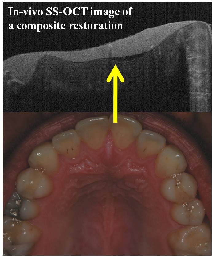
Dental hard tissues and biomaterials can be non-destructively assessed using optical coherence tomography (OCT), an emerging diagnostic tool. Swept Source (SS)-OCT has an improved imaging resolution and speed. Data obtained in B-scans can be used to assess the quality and depth-resolved information on the tissues. We are working to develop new methods and devices to take advantage of SS-OCT both in the research and the practice of cariology and operative dentistry, in collaborative projects.
 © Cariology and
Operative Dentistry
© Cariology and
Operative Dentistry